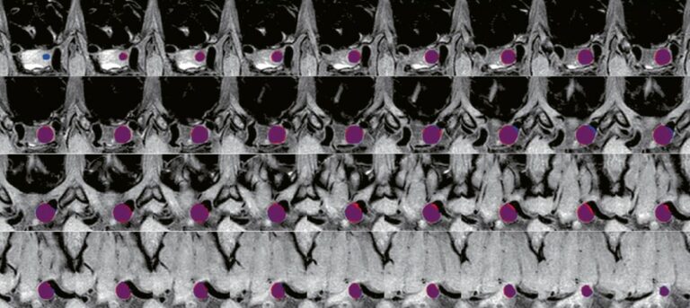


Knee landmarks detection via deep learning
A deep learning-based approach was developed and validated in this study which aimed to automatically measure the patellofemoral instability (PFI) indices related to patellar height and trochlear dysplasia in knee MRI scans. The authors included a total of 763 knee MRI slices from 95 patients, annotating 3,393 anatomical landmarks. The results indicated that the developed models achieved good accuracy in